Enter R180 in the search box to learn more about "Surgery for Retinal Detachment: What to Expect at Home". This can occur in or around the eye and lead to permanent vision loss. If the retina cannot be reattached, the eye will continue to lose sight and ultimately become blind. Laser surgery is one way. Accessed Sept. 12, 2018. You will need to be careful not to get any soap or water in your eye. Air, gas or silicone oil is then injected into the vitreous space to help flatten the retina. How quickly the surgery needs to be done depends on the location and extent of the detachment. 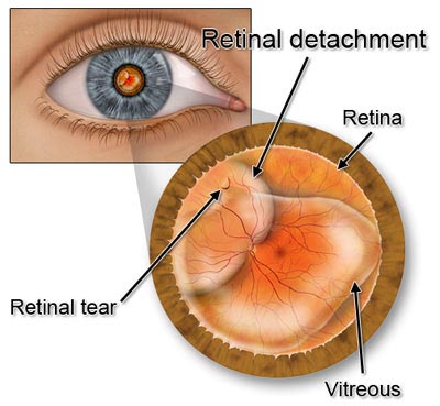 1 in 1000 risk of bleeding. It is important to maintain this position so the gas bubble pushes the retina into place. Simple annoyance or the sign of a problem?
After the surgery, your eye may feel a little sore. Your final visual outcome will depend on how much nerve damage occurred with your retinal detachment. You need to keep in mind that our goal is to maximize the vision in your affected eye. Your doctor then injects a small amount of intraocular gas into the vitreous. This scar tissue seals the tear or helps the retina reattach to the underlying tissues and keeps it in the correct place. National Eye Institute. If the macula was involved for less than 1 week, vision will usually be improved, but not to 20/20 (normal). Doctors determine the type of surgery needed based on several factors, including the location and size of the detachment and whether the person has had cataract surgery. Diseases of the visual system. Further Information But each person recovers at a different pace. This includes moving quickly, lifting anything heavy, or doing activities such as cleaning or gardening. After the surgery you will have an eye shield placed on your eye. The eye doctor injects a bubble of gas into the eye. You may have to wear an eye patch or shield for a few days. The band pushes gently on the sides of your eye and moves them inward toward your retina, which helps your retina reattach. Your eye doctor may put drops in your eye to prevent infection and keep the pupil from opening wide or closing. Patients often complain of flashes, new floaters and a shadow forming in their vision when a retinal detachment occurs. After surgery, it may take several weeks or months to regain full vision in the affected eye. Pneumatic retinopexy (noo-mat-ick RET-ih-no-pek-see), In pneumatic retinopexy, your doctor will inject a small air bubble into your eye. Have any of your family members ever had a retinal detachment? The biggest reason for failure of retinal detachment surgery is the formation of scar tissue that redetaches the retina (proliferative vitreoretinopathy). Injecting air or gas into your eye. Various techniques are available. Check out these best-sellers and special offers on books and newsletters from Mayo Clinic Press. If you take aspirin or some other blood thinner, ask your doctor if and when to start taking it again. The buckle is placed in a way that doesn't block your vision, and it usually remains in place permanently. Posterior Vitreous Detachment: Vision Problems as You Age, Failure to repair the detached retina, which can mean more surgery. Laser photocoagulation and cryotherapy may also be used in conjunction with surgery for complete treatment. Be sure to make and go to all appointments, and call your doctor or nurse advice line (811 in most provinces and territories) if you are having problems. The symptoms described above may not necessarily mean that you have a detached retina. You may have to wear a patch or shield over the eye for a day or more. Retinal detachment happens when your retina (a light-sensitive layer of tissue at the back of your eye) is pulled away from its normal position. Opens in a new window. As with laser photocoagulation, your doctor may recommend that you rest after the procedure so the scars can form and your eye can heal. Retinal detachment is a medical emergency, and early treatment isimportant to protect your vision. Failure to repair the retina always results in loss of vision to some degree. A special intraocular gas may be injected into the eye, creating a bubble that expands and pushes the retina against the back of the eye. If the macula detaches, it is too late to restore normal vision. What is a detached retina and what are the causes? Do not lie on your back. https://www.aao.org/eye-health/diseases/detached-torn-retina. Insert a tiny needle into your eye and remove a small amount of fluid, Inject a small amount of air into your eye, Use laser or freeze treatment to repair any holes or tears in your retina, Hold your head in a certain position for several days to keep the air bubble in the right spot, Avoid some activities like flying in an airplane, intense exercise, and heavy lifting while your eye heals, Have a follow-up visit with your doctor to make sure your eye is healing, Wear a patch over your eye for about a day, Avoid some activities like heavy lifting or heavy exercise while your eye heals, Funding for Training and Career Development, Diversity, Equity, Inclusion and Accessibility at NEI, Learn more about laser surgery and freeze treatment. The buckle stays in your eye permanently. The retina sends visual images to the brain through the optic nerve. Risks and complications of all retinal surgeries include bleeding and infection. Your surgeon will review appropriate head positioning with you and your family members after surgery. Redness and swelling in your leg or groin. The sooner the detachment is repaired, the sooner the rods and cones will begin to recover. The retina is the light-sensitive tissue in the back of the eye.
2nd ed. You stand the best chance at a good outcome if the repair is done before the center part of the retina, called the macula, detaches. In: Ferri's Clinical Advisor 2019. Read our updated information about wearing a mask for your visit, and our visitor policy. The vitreous is the clear collagen gel that fills the eye between the retina and the lens. 2005 - 2022 WebMD LLC. Usually the vitreous separates from the retina without causing a problem. Follow the steps below to get better as quickly as possible. Your ophthalmologist will prescribe any necessary medications for you and advise you when to resume normal activity. Your doctor also uses cryopexy during the procedure to repair the retinal break. Your eye surgeon may need to freeze several areas before the tear is sealed or the retina is reattached. Most of the time, the retina can be reattached with one operation. If the doctor gave you a prescription medicine for pain, take it as prescribed. You will also get instructions about taking any new medicines. Most retinal detachment surgery is successful, although a second operation is sometimes needed. Airplane travel is dangerous. Have you had any symptoms in your other eye? Learn more about the COVID-19 vaccine. People with a gas bubble in the eye may not fly or go to high altitudes until the gas bubble dissolves. Your doctor may also use a laser or freeze treatment to repair any tears in your retina. If you have aretinaldetachment,you may need surgery to reattach your retinato the back of your eye within a few days. This procedure takes about one hour.
National Institutes of Health/National Eye Institute, Facts About Retinal Detachment., National Library of Medicine, Medline Plus, Retinal detachment repair.. What's the most likely cause of my symptoms? . If the retina has just started to detach, a procedure called pneumatic retinopexy may be done to repair it. Retinal detachments DO NOT get better without treatment. More than 9 out of 10 detachments can be repaired. Both of these procedures are done on an outpatient basis. Most people can go home the same day, but youll need someone to drive you home. What are other possible causes of my symptoms? Ask your ophthalmologist about the risks and benefits of your treatment options. Opens in a new window. The retina normally lies smoothly and firmly against the inside back wall of the eyeball and functions much like the film in the back of a camera. digital privacy statement. What will determine whether I should plan for a follow-up visit? browse our specialists. You are then positioned so the gas bubble floats up against the hole in the retina and pushes it back into place. After the vitreous is removed, your doctor may treat the retina with photocoagulation or cryotherapy to seal the tear. However, once the retina has detached, the photoreceptors may never recover completely. In: Walls RM, Hockberger RS, Gausche-Hill M, eds. Although your vision may not return completely to its previous state, the goal of surgery is to restore usable vision. As a result of injury, tumors, or disease, the retina can become completely or partially detached causing diminished vision. Your vision after surgery will likely be worse if your macula has become detached. Vitrectomy may be combined with a scleral buckling procedure. Your eye surgeon will decide which procedure for retinal detachment is right for you. In: The Retinal Atlas. Peripheral retinal degenerations and rhegmatogenous retinal detachment. If any part of the retina is lifted or pulled from its normal position, it is considered detached and will cause some vision loss. The laser is then focused over the retinal tear or small detachment. The vitreous is replaced by natural fluid produced inside the eye. 6th ed. Learn more about our research and professional education opportunities. NYU Langone Health is one of the nations premier academic medical centers. You may drive when your vision allows it. Do not remove this shield until we see you in the clinic the following day when we will remove it for you. Your eye produces fluid that eventually replaces the gas and fills the eye. In certain cases we may use silicone oil instead of gas; your surgeon will review with you if this is appropriate for your surgery. If you have some pain we recommend you take Acetaminophen (Tylenol). The retina is the light-sensitive layer of nerve tissue that lines the inside of the eye and sends visual messages through the optic nerve to the brain. In most cases, the procedures do not require an overnight hospital stay.
Doctors use this method, called pneumatic retinopexy, if the retina has just begun to detach. Sterile technique is used during the procedure to minimize risk of infection. The type of surgery your surgeon recommends will depend on several factors, including how severe the detachment is. It also will increase the chance of preserving good vision. Any surgery has risks; however, an untreated retinal detachment will usually result in permanent severe vision loss or blindness. The laser emits a beam of light that travels through the eye and burns the area around the retinal tear or detachment to create a scar. As we get older, the vitreous may pull away from its attachment to the retina at the back of the eye. Ophthalmology. This procedure indents the wall of the eye and relieves some of the force caused by the vitreous tugging on the retina. Conditions that can increase the chance of a retinal detachment include nearsightedness; previous cataract surgery; glaucoma; severe trauma; previous retinal detachment in your other eye; family history of retinal detachment; or weak areas in your retina that can be seen by your ophthalmologist. If positioned properly, the bubble pushes the area of the retina containing the hole or holes against the wall of the eye, stopping the flow of fluid into the space behind the retina. However, in some people, there may be a recurrence of retinal detachment that may require two or more surgeries to treat. The retina can be repaired by laser, cryoprobe, or surgery. It is typically performed the under local anesthesia so that you are awake and comfortable during the procedure and have minimal complications from anesthesia postoperatively. The retina is a thin layer of tissue in the back of the eye that is crucial for vision. Learn about causes, symptoms, and treatments. Often, it will be less than 20/200, the limit for legal blindness. This scar tissue helps seal the tear or reattach a detached portion of retina to underlying tissue. Editorial team. In: Goldman L, Schafer AI, eds. https://www.asrs.org/patients/retinal-diseases/6/retinal-detachment. This can help prevent further detachment of the retina. American Society of Retina Specialists. Philadelphia, PA: Elsevier; 2018:chap 109. Do they require any special preparation? Follow us on LinkedIn. Gariano, R. American Family Physician, April 1, 2004. For this procedure, the doctor will numb your eye and then put a small, freezing probe on it. Your doctor discusses anesthesia options with you before surgery.
1 in 1000 risk of bleeding. It is important to maintain this position so the gas bubble pushes the retina into place. Simple annoyance or the sign of a problem? After the surgery, your eye may feel a little sore. Your final visual outcome will depend on how much nerve damage occurred with your retinal detachment. You need to keep in mind that our goal is to maximize the vision in your affected eye. Your doctor then injects a small amount of intraocular gas into the vitreous. This scar tissue seals the tear or helps the retina reattach to the underlying tissues and keeps it in the correct place. National Eye Institute. If the macula was involved for less than 1 week, vision will usually be improved, but not to 20/20 (normal). Doctors determine the type of surgery needed based on several factors, including the location and size of the detachment and whether the person has had cataract surgery. Diseases of the visual system. Further Information But each person recovers at a different pace. This includes moving quickly, lifting anything heavy, or doing activities such as cleaning or gardening. After the surgery you will have an eye shield placed on your eye. The eye doctor injects a bubble of gas into the eye. You may have to wear an eye patch or shield for a few days. The band pushes gently on the sides of your eye and moves them inward toward your retina, which helps your retina reattach. Your eye doctor may put drops in your eye to prevent infection and keep the pupil from opening wide or closing. Patients often complain of flashes, new floaters and a shadow forming in their vision when a retinal detachment occurs. After surgery, it may take several weeks or months to regain full vision in the affected eye. Pneumatic retinopexy (noo-mat-ick RET-ih-no-pek-see), In pneumatic retinopexy, your doctor will inject a small air bubble into your eye. Have any of your family members ever had a retinal detachment? The biggest reason for failure of retinal detachment surgery is the formation of scar tissue that redetaches the retina (proliferative vitreoretinopathy). Injecting air or gas into your eye. Various techniques are available. Check out these best-sellers and special offers on books and newsletters from Mayo Clinic Press. If you take aspirin or some other blood thinner, ask your doctor if and when to start taking it again. The buckle is placed in a way that doesn't block your vision, and it usually remains in place permanently. Posterior Vitreous Detachment: Vision Problems as You Age, Failure to repair the detached retina, which can mean more surgery. Laser photocoagulation and cryotherapy may also be used in conjunction with surgery for complete treatment. Be sure to make and go to all appointments, and call your doctor or nurse advice line (811 in most provinces and territories) if you are having problems. The symptoms described above may not necessarily mean that you have a detached retina. You may have to wear a patch or shield over the eye for a day or more. Retinal detachment happens when your retina (a light-sensitive layer of tissue at the back of your eye) is pulled away from its normal position. Opens in a new window. As with laser photocoagulation, your doctor may recommend that you rest after the procedure so the scars can form and your eye can heal. Retinal detachment is a medical emergency, and early treatment isimportant to protect your vision.
Failure to repair the retina always results in loss of vision to some degree. A special intraocular gas may be injected into the eye, creating a bubble that expands and pushes the retina against the back of the eye. If the macula detaches, it is too late to restore normal vision. What is a detached retina and what are the causes? Do not lie on your back. https://www.aao.org/eye-health/diseases/detached-torn-retina. Insert a tiny needle into your eye and remove a small amount of fluid, Inject a small amount of air into your eye, Use laser or freeze treatment to repair any holes or tears in your retina, Hold your head in a certain position for several days to keep the air bubble in the right spot, Avoid some activities like flying in an airplane, intense exercise, and heavy lifting while your eye heals, Have a follow-up visit with your doctor to make sure your eye is healing, Wear a patch over your eye for about a day, Avoid some activities like heavy lifting or heavy exercise while your eye heals, Funding for Training and Career Development, Diversity, Equity, Inclusion and Accessibility at NEI, Learn more about laser surgery and freeze treatment. The buckle stays in your eye permanently. The retina sends visual images to the brain through the optic nerve. Risks and complications of all retinal surgeries include bleeding and infection. Your surgeon will review appropriate head positioning with you and your family members after surgery. Redness and swelling in your leg or groin. The sooner the detachment is repaired, the sooner the rods and cones will begin to recover. The retina is the light-sensitive tissue in the back of the eye. 2nd ed. You stand the best chance at a good outcome if the repair is done before the center part of the retina, called the macula, detaches. In: Ferri's Clinical Advisor 2019. Read our updated information about wearing a mask for your visit, and our visitor policy. The vitreous is the clear collagen gel that fills the eye between the retina and the lens. 2005 - 2022 WebMD LLC. Usually the vitreous separates from the retina without causing a problem. Follow the steps below to get better as quickly as possible. Your ophthalmologist will prescribe any necessary medications for you and advise you when to resume normal activity. Your doctor also uses cryopexy during the procedure to repair the retinal break. Your eye surgeon may need to freeze several areas before the tear is sealed or the retina is reattached. Most of the time, the retina can be reattached with one operation. If the doctor gave you a prescription medicine for pain, take it as prescribed. You will also get instructions about taking any new medicines. Most retinal detachment surgery is successful, although a second operation is sometimes needed. Airplane travel is dangerous. Have you had any symptoms in your other eye? Learn more about the COVID-19 vaccine. People with a gas bubble in the eye may not fly or go to high altitudes until the gas bubble dissolves. Your doctor may also use a laser or freeze treatment to repair any tears in your retina. If you have aretinaldetachment,you may need surgery to reattach your retinato the back of your eye within a few days. This procedure takes about one hour.
National Institutes of Health/National Eye Institute, Facts About Retinal Detachment., National Library of Medicine, Medline Plus, Retinal detachment repair.. What's the most likely cause of my symptoms? . If the retina has just started to detach, a procedure called pneumatic retinopexy may be done to repair it. Retinal detachments DO NOT get better without treatment. More than 9 out of 10 detachments can be repaired. Both of these procedures are done on an outpatient basis. Most people can go home the same day, but youll need someone to drive you home. What are other possible causes of my symptoms? Ask your ophthalmologist about the risks and benefits of your treatment options. Opens in a new window. The retina normally lies smoothly and firmly against the inside back wall of the eyeball and functions much like the film in the back of a camera. digital privacy statement. What will determine whether I should plan for a follow-up visit? browse our specialists. You are then positioned so the gas bubble floats up against the hole in the retina and pushes it back into place. After the vitreous is removed, your doctor may treat the retina with photocoagulation or cryotherapy to seal the tear. However, once the retina has detached, the photoreceptors may never recover completely. In: Walls RM, Hockberger RS, Gausche-Hill M, eds. Although your vision may not return completely to its previous state, the goal of surgery is to restore usable vision. As a result of injury, tumors, or disease, the retina can become completely or partially detached causing diminished vision. Your vision after surgery will likely be worse if your macula has become detached. Vitrectomy may be combined with a scleral buckling procedure. Your eye surgeon will decide which procedure for retinal detachment is right for you. In: The Retinal Atlas. Peripheral retinal degenerations and rhegmatogenous retinal detachment. If any part of the retina is lifted or pulled from its normal position, it is considered detached and will cause some vision loss. The laser is then focused over the retinal tear or small detachment. The vitreous is replaced by natural fluid produced inside the eye. 6th ed. Learn more about our research and professional education opportunities. NYU Langone Health is one of the nations premier academic medical centers. You may drive when your vision allows it. Do not remove this shield until we see you in the clinic the following day when we will remove it for you. Your eye produces fluid that eventually replaces the gas and fills the eye. In certain cases we may use silicone oil instead of gas; your surgeon will review with you if this is appropriate for your surgery. If you have some pain we recommend you take Acetaminophen (Tylenol). The retina is the light-sensitive layer of nerve tissue that lines the inside of the eye and sends visual messages through the optic nerve to the brain. In most cases, the procedures do not require an overnight hospital stay.
Doctors use this method, called pneumatic retinopexy, if the retina has just begun to detach. Sterile technique is used during the procedure to minimize risk of infection. The type of surgery your surgeon recommends will depend on several factors, including how severe the detachment is. It also will increase the chance of preserving good vision. Any surgery has risks; however, an untreated retinal detachment will usually result in permanent severe vision loss or blindness. The laser emits a beam of light that travels through the eye and burns the area around the retinal tear or detachment to create a scar. As we get older, the vitreous may pull away from its attachment to the retina at the back of the eye. Ophthalmology. This procedure indents the wall of the eye and relieves some of the force caused by the vitreous tugging on the retina. Conditions that can increase the chance of a retinal detachment include nearsightedness; previous cataract surgery; glaucoma; severe trauma; previous retinal detachment in your other eye; family history of retinal detachment; or weak areas in your retina that can be seen by your ophthalmologist. If positioned properly, the bubble pushes the area of the retina containing the hole or holes against the wall of the eye, stopping the flow of fluid into the space behind the retina. However, in some people, there may be a recurrence of retinal detachment that may require two or more surgeries to treat. The retina can be repaired by laser, cryoprobe, or surgery. It is typically performed the under local anesthesia so that you are awake and comfortable during the procedure and have minimal complications from anesthesia postoperatively. The retina is a thin layer of tissue in the back of the eye that is crucial for vision. Learn about causes, symptoms, and treatments. Often, it will be less than 20/200, the limit for legal blindness. This scar tissue helps seal the tear or reattach a detached portion of retina to underlying tissue. Editorial team. In: Goldman L, Schafer AI, eds. https://www.asrs.org/patients/retinal-diseases/6/retinal-detachment. This can help prevent further detachment of the retina. American Society of Retina Specialists. Philadelphia, PA: Elsevier; 2018:chap 109. Do they require any special preparation? Follow us on LinkedIn. Gariano, R. American Family Physician, April 1, 2004. For this procedure, the doctor will numb your eye and then put a small, freezing probe on it. Your doctor discusses anesthesia options with you before surgery.